Patients
-
- Angiography
- Angioplasty and Stenting
- Aortic Aneurysms
- Biliary Drainage and Stenting
- Carotid Artery Stenting
- Central Venous Access
- Colonic Stenting
- Fibroids
- Gastrointestinal Haemorrhage
- Gastrostomy
- Hepatic Malignancies
- Kidney Tumour Ablation
- Minimally Invasive Treatments for Vascular Disease
- Nephrostomy
- Oesophageal Stents
- Pelvic Venous Congestion Syndrome
- Percutaneous Nephrolithotomy
- Prostate Artery Embolisation PAE
- Pulmonary Arteriovenous Malformations
- PAE Patient Information Leaflet
- Ureteric Stenting
- Varicoceles
- Varicose Veins
- Vascular Malformations
- Vertebral Compression Fractures
- Vertebroplasty and Kyphoplasty
What is Interventional Radiology?
Interventional radiology (IR) is one of the most innovative and fastest-growing specialities in the field of medicine. An interventional radiologist is a doctor who performs image-guided procedures, fully interprets the imaging required to guide and monitor a patient’s response to those procedures and provides pre and post procedural care for our patients.
Interventional radiologists have been developing innovative alternatives to open surgery since the 1960s, but it has really been in the last 20 years that we have seen major advances in what we can achieve, with the development of modern imaging and operative technologies. These allow IRs to perform the most complex of tasks through a small pinpick in the skin, advancing catheters, guidewires or needles to targets deep inside the body, while avoiding damage to critical surrounding tissues.
Interventional radiology operating theatres contain imaging equipment such as ultrasound machines, X-ray machines and CT scanners so that we can monitor the operation in real time.
Because of this keyhole approach, IR procedures are often termed ‘minimally invasive’ and rightly so. It means patients usually heal from their operation far more speedily, have no obvious scar and can often have pretty complex procedures performed without having to stay longer than a few hours in hospital, meaning that patients can get back to their normal lives more quickly.
IR targets many of today’s toughest medical problems, including vascular disease, many types of cancer and men’s and women’s health conditions with minimal discomfort and unprecedented precision. IR’s culture of continuous innovation has led to development of targeted treatments for virtually every organ system in the body. Innovative treatments developed by interventional radiologists are now used routinely across all fields of medicine.
This short film shows what interventional radiology is:
How is it done?
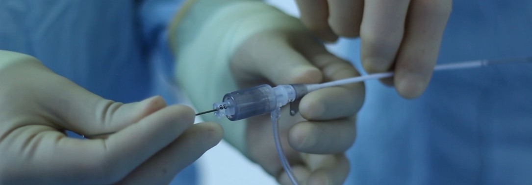
There are different techniques depending on the procedure but put broadly we access the natural routes in the body if at all possible. This is particularly suitable for the various tubes in the body such as blood vessels, bowel, and urinary tract. We use an accessible entry point and with the help of catheters and other medical devices travel in the vessel to the point of disease. As we are travelling within the natural passages of the body, there is less pain and trauma.
What are the advantages of Interventional Radiology?
Interventional Radiology is much less invasive than conventional treatments and most procedures will be done through incisions less than 5mm. It is often possible to avoid general anaesthetics and the majority of our procedures are performed under local anaesthesia sometimes combined with sedation. This means patients can recover faster, often need less time off work and the cost of procedures are reduced.
What treatments can be undertaken with Interventional Radiology?
The range of conditions which can be treated by IR is enormous and continually expanding. Here are some well known conditions which can be treated using IR:
Blood vessel disease
Arteries
Narrowing of arteries leading to restricted blood flow (peripheral vascular disease): Interventional radiologists treat this by using balloons to stretch the vessel (balloon angioplasty, PTA) and sometimes metal springs called stents to hold them open. Sometimes arteries or bypass grafts block suddenly with a rapid loss of blood supply to the limb. Unless the blood supply is restored this can lead to amputation. Interventional radiologists can help by infusion of clot busting drugs directly into the artery via small catheters thus saving many limbs.
Expanded arteries (aneurysms) at risk of rupture and bleeding: IRs treat these by relining the vessel with a tube called a stent graft
Bleeding (haemorrhage). This is the most common vascular emergency treated by IR. Haemorrhage can come from almost anywhere e.g. from the gut, secondary to major injury or following birth. Bleeding can often permanently be stopped by blocking the vessel (embolization), relining the vessel with a stent graft or by blowing up a balloon in the vessel to stop the bleeding until emergency surgery can be performed. Interventional radiology is also used to prevent bleeding during some sorts of surgery e.g. during caesarean section in patients with a high risk of bleeding from an abnormal placenta (post partum haemorrhage).
Veins
Blood clots in the lung (pulmonary embolism, PE): interventional radiologists perform 2 different forms of treatment, placement of devices (inferior vena cava filters) to capture blood clots before they reach the lung preventing further PE. When there is a massive PE causing collapse an interventional radiologist may use small catheter tubes to break up the blood clot and restore blood flow.
Dilated veins (varicose veins): these most commonly occur in the legs but can occur in the pelvis or scrotum. These can be treated by blocking the vein by heat treatment (laser or microwave) or by the use of irritant drugs and embolisation techniques.
Blocked veins: this can occur in the context of blood clot in the veins (venous thrombosis, DVT) which is sometimes treated by the injection of blood clot dissolving medicines (thrombolysis) through a small catheter passed into the vein. Some patients develop blood clots as a result of a narrowing in a vein, when the clot has been broken down using balloons and stents. Sometimes tumours in the chest will compress a vein leading to facial swelling, headache and other symptoms which can usually be relieved with a stent.
Non vascular intervention
This is sometimes referred to as interventional oncology but the treatments are also effective in benign conditions. IR therapies are used for the following:
- to treat the tumour / cancer (tumour ablation, embolization)
- to relieve the effects of the cancer on other systems e.g. blockage of the gullet (oesophagus), bowel, kidney (nephrostomy) or liver (biliary drainage)
- To drain collections of fluid or pus in the chest or abdomen
- To place feeding tubes (gastrostomy, jejunostomy)
- To treat collapsed spinal bones (vertebroplasty)
Tumour therapies: these treatments are intended to shrink or destroy tumours at their primary site or which have spread to other areas (metastases). This is an area of increasing interest and leading to improved survival with reduced morbidity.
Liver, kidney and other tumours (e.g. bone, lung): these can be treated by destructive therapies (ablation) usually involving heat (radiofrequency, laser, microwave, ultrasound) or cold damage (cryotherapy). The treatment is performed and monitored using imaging (ultrasound, computed tomography or magnetic resonance imaging).
Uterine fibroids : heavy menstrual bleeding and pain can be caused by benign tumours called fibroids. These can be treated by blocking blood vessels (uterine fibroid embolization, UFE) which leads to shrinkage. Embolization is sometimes combined with drug therapy (chemoembolization) or radiotherapy (radioembolization) which targets the effect to the tumour and limits some of the side effects of cancer therapy.
Stone Disease
Kidney stones are not uncommon and cause pain, infection and blockage of the kidney. Obstruction of the kidney in the presence of infection will rapidly cause irreversible kidney damage. Interventional techniques include placing a tube in the kidney (nephrostomy) to allow the urine to drain and removing the stones using a variety of instruments placed through the skin into the kidney. Large kidney stones are best dealt with by creating a tunnel into the kidney through a small skin incision and then passing an endoscope directly into the kidney, breaking the stone with special instruments and pulling the fragments out (percutaneous nephrolithotomy).
Gall stones are one of the most common upper abdominal disorders. Most are dealt with by laparoscopic surgery. When stones or tumour stop bile draining from the liver this causes jaundice, this is usually treated via a telescope passed down the throat (endoscopy) but sometimes requires an interventional radiologist to perform drainage by placing catheter tubes through the liver to either remove the stones or place stents to allow drainage.